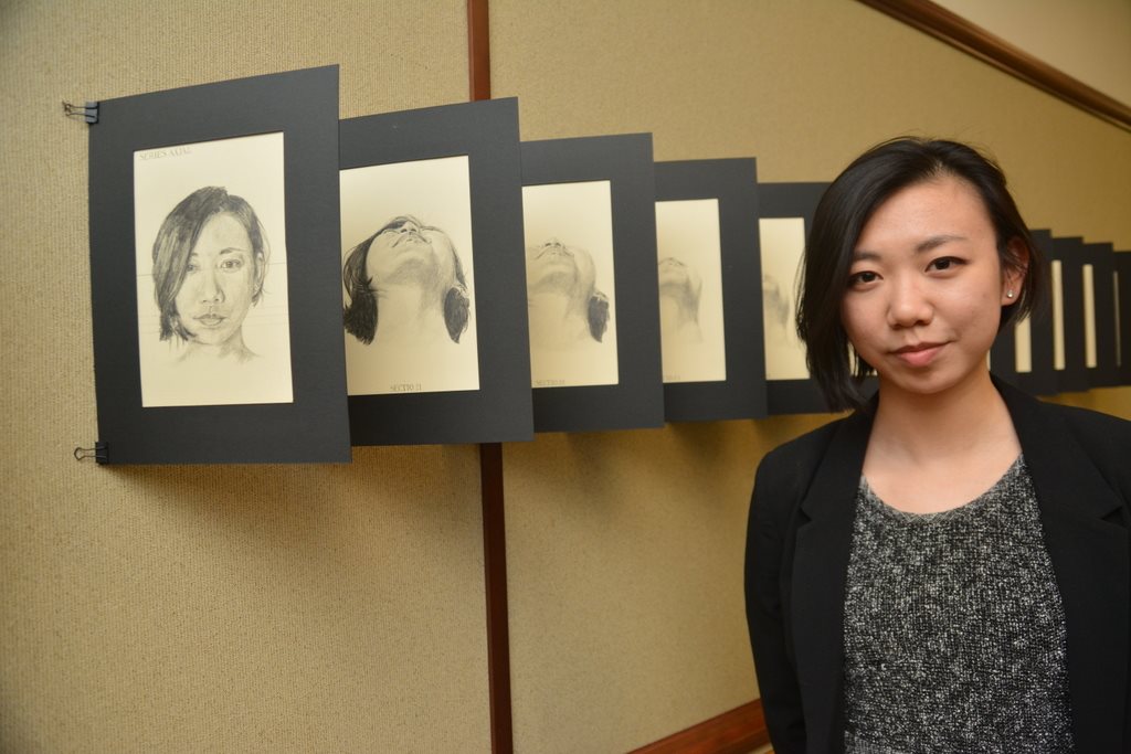
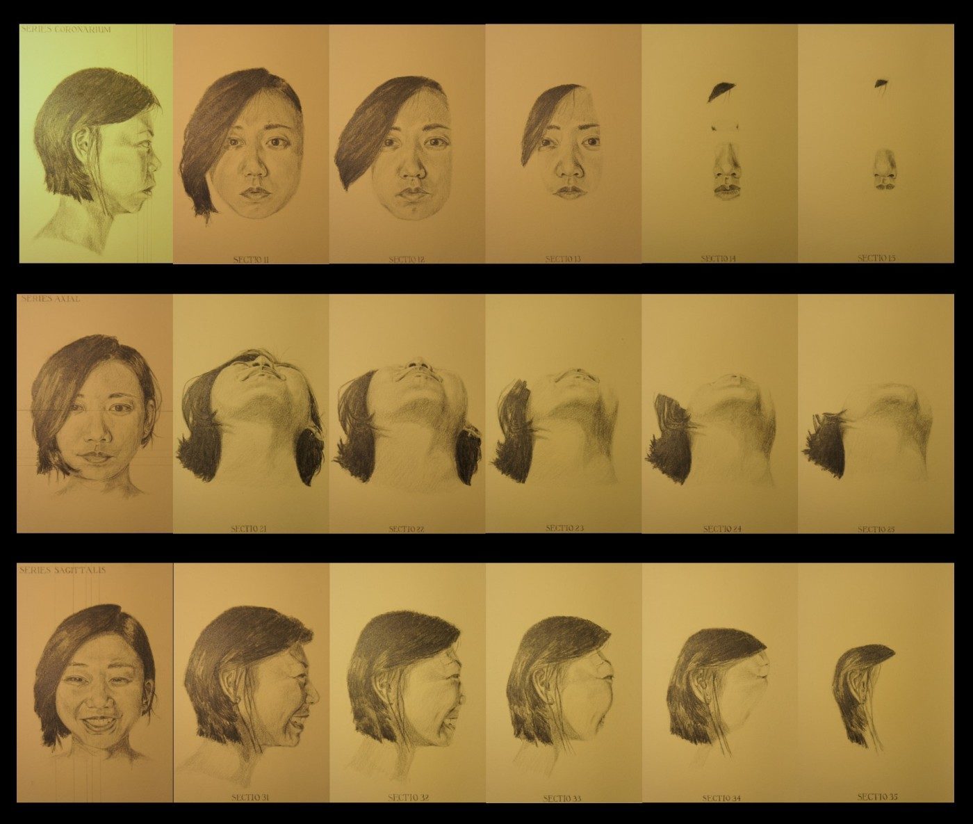
Atlas
Siyu Xiao (2015)
graphite on paper

Photo credit: Photograph by John Curtis at Yale Medicine.
The idea for this piece came to me in the last few weeks of my first-year anatomy class. I wanted to create a work for my school’s annual Service of Gratitude, where the class commemorates those individuals who donated their bodies for our education.
I won’t say too much about the piece, as I’d like to allow the viewer his or her own experience. I will just say that learning about the human body for the first time through gross “sections,” radiographic “slices” and illustrated muscle groups in various atlases came with a bizarre, inhuman — or inhumane, even — feeling to it. I could not stop thinking about how learning the human body meant that I had to study it in its most mutilated forms. It was just too ironic.
At the same time, the beauty and diligence with which anatomists and pathologists prepared those sections for our education was moving. I was particularly drawn to a set of brain stem axial images, blown up to many times their original size, printed on sturdy vintage-looking panels and annotated in stately serif font. Historical but elegant, much more so than most of the modern pedagogical materials I encounter.
The image above is a rough stitch of 18 JPEGs combined into one file, which I did in PowerPoint. As the viewer walks past each drawing, one by one, through the three series, the viewer is able to appreciate the progression and expansion of the piece. The first drawing in each series is labeled “Series Coronarium,” “Series Axial” or “Series Sagittalis.” The following drawings are labeled “Section #” from 1-5.

