
He had come in overnight, already intubated and unresponsive when we met him. Here was a man, bits and pieces of him lost over the years, after many difficult battles with many difficult infections. As we softly peeled back the rolls of gauze around his leg, the chief surgery resident gagged behind his mask and turned away. What might have started out as a regular case of uncontrolled diabetes or vascular disease spiraled into necrotizing fasciitis, the infamous flesh-eating disease.
The patient’s infection crept up his thigh as the day crept on. By that afternoon, I stood in the stark operating room, rocking back and forth on my own toes while hoisting the decaying leg of a man I still had yet to meet. The only way to get ahead of necrotizing fasciitis is source control, meaning we needed to cut off the infection upstream of the source. In a word: amputation.
I clutched the edematous flesh of the patient’s calf with my left hand, gripping his gangrenous foot against my chest with my right hand. There was no blood supply left to that rotting leg, hardly enough to heal an amputated limb. The surgery intern stood next to me, suctioning, suctioning, suctioning, as the attending surgeon peeled and peeled and peeled at the fascia. The surgeon scraped away what he could and told us to pray for a miracle.
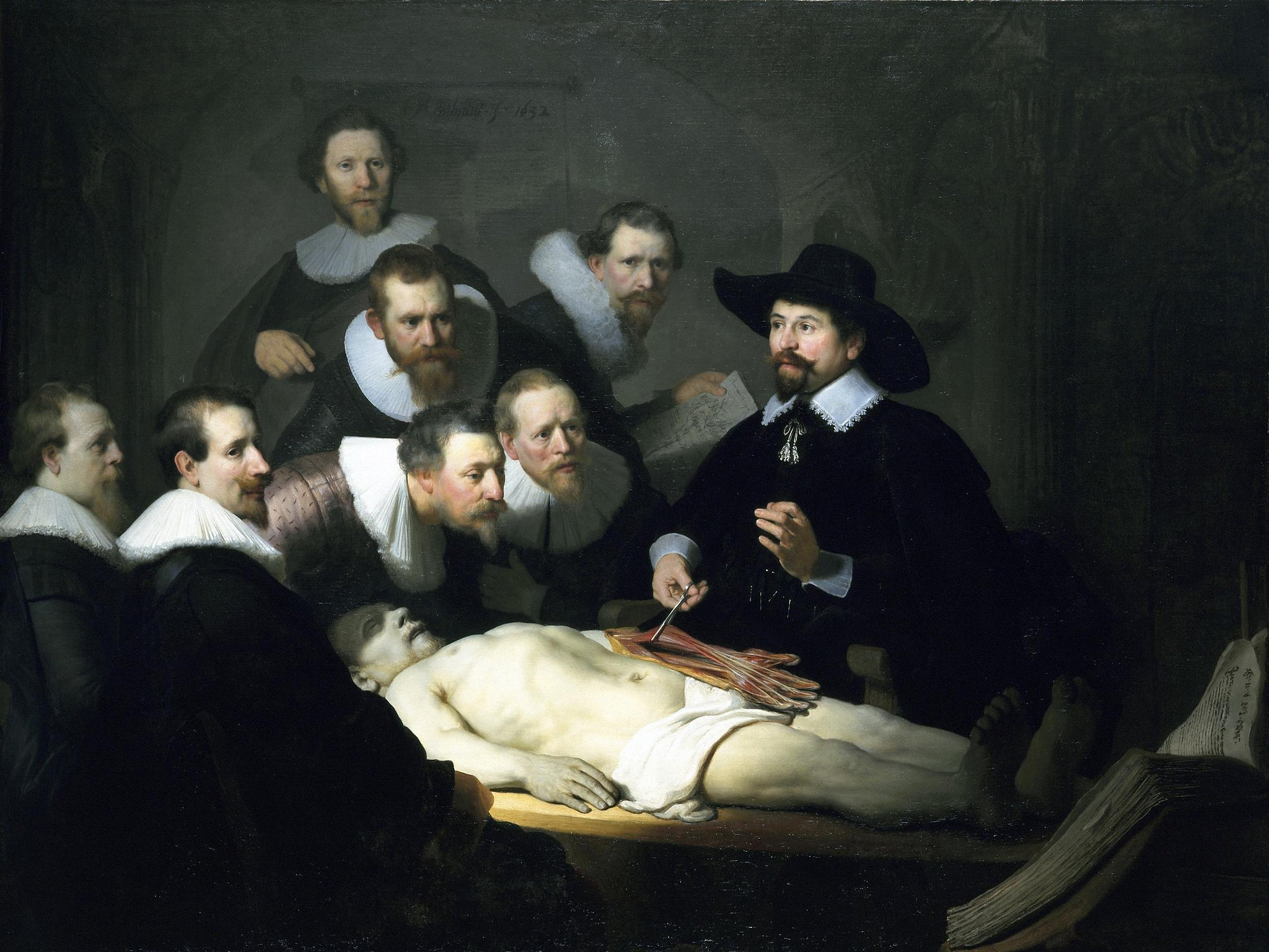
There were seven of us standing around the table as the attending surgeon debrided the infected fascia. The vascular surgeon came in the room and barked at us to identify the structures before us. “What’s that artery?” he interrogated us. “I’ll give you a hint,” he said, “there’s a deep and a superficial.” We named the sural nerve and iliotibialis band and the great saphenous vein. As we clamored around the table, I suddenly thought of the Rembrandt painting: The Anatomy Lesson of Dr. Nicolaes Tulp.
In the painting, the Dutch master depicts an anatomy lesson given by a surgeon, Dr. Nicolaes Tulp. Several individuals, most likely other doctors, are crowded around the table observing a corpse laying exposed with an arm flayed open. The painting is a famous one, a painting that I have seen many times, from European history classes on the Dutch Golden Age to preclinical lectures on brachial plexus injuries. As I left the operating room that day, I thought briefly of the tableau we had made as we held onto our patient, the memory suspended in my mind as a painting on the wall.
The day after our anatomy lesson, the chief resident called me back into the operating room. Knowing the odds were stacked against them, the patient and his husband had asked for one more trip to the table. They were still praying for that miracle, that the infection might somehow clear without any blood to clear it, that an amputation might somehow heal without any blood to heal it. And so I stood in the operating room once more, waiting in apprehension.
We set the stage, raised the drapes, focused the lights as we had done for him the day before. “The anatomy really is quite stunning,” the attending surgeon remarked as we began the dissection. He pointed to muscle after muscle, asking me to identify the adductor magnus and longus, the sartorius and gracilis, the vastus medius, the way I would on a cadaver. I held a blue plastic tube that suctioned what was left of this man’s blood from what was left of his body. We worked intensely for only a few minutes, before the surgeon himself lifted the patient’s leg.
He paused. There, we saw, was the vast spread of infection, even farther, even deeper than the day before. The surgeon dissected the leg muscles with the same precision that I imagine Dr. Nicolaes Tulp had. He separated the gray, rotting fascia, pulling it apart with the ease that could be found on a corpse, quizzing me on each layer all the while. He put his blade down. “We’re finished,” he told the room. “I’m not going to cut this man anymore.” We quietly removed all the drapes and wrapped the patient in soft gauze once again. I never saw him again, but I heard from the nursing staff that he died before the next day. His husband was at his side, weeping with grief and relief now that it was all over.
—
That surgery was many months ago and that patient has been gone for as long, but I have looked at Rembrandt’s painting and thought of that anatomy lesson a hundred times since then without really knowing why it remains rooted in my mind. There are so many scenes in the hospital that are not refined, that are not composed, that are not worth hanging in a gallery with pride. The hospital is not beautiful. It is tiresome and stark, with long hours and cracked linoleum tiles. And yet I still have an image of all of us arranged around this patient just as Rembrandt would have arranged us.
A man came in with an infection and became a body for an anatomy lesson. Death appeared inevitable for this patient, so the focus shifted from saving his life to learning the aspects of his case that might prevent death in another, more fortunate patient. That anatomy lesson I received in the operating room was the last frontier that medicine could reach. In the throes of death, a man became a body even before he died. The most that medicine could extract from this life was one more anatomy lesson, one more opportunity for the education of a young scientist in the crusade against death.
In medical school we do not learn much about death, but rather the tools and tricks that a doctor must know to stave off death. In the fight against death, we sometimes fail to recognize the things that make up a life. Patients enter the hospital and become diagnoses, statistics and risk factors. In preserving vital signs and organ function, we may sacrifice the joy of dancing or the delight of a home-cooked meal. We do not realize the boundaries of our abilities until a patient dies despite our best efforts. For all the data-driven empathy training and peer-reviewed articles about grief, science is not able to restore the soul.
Death is, in many ways, a failure of medicine. But what if dying and death could be as beautiful and revered as a painting? Art has an extraordinary way of elevating tragedies, of glamorizing treason and plague and sorrow. In every catastrophe, there is composition, color and chiaroscuro. Scenes of low spirits become grand masterpieces of resilience and fortitude. Medicine fears death and fights it; art can revere death and celebrate it.
The Anatomy Lesson transforms the body into more than just a prop or a teaching aid. Rembrandt’s painting returns the body to its higher purpose. He depicts the body as the divine form that it is, and the great privilege it is to study such a form. Through art, we can reach beyond death, recapturing the beauty and humanity in the horrible things we must do and see in medicine. Art restores the humanity that patients lose when they come to the hospital and become bodies rather than people. Paintings like The Anatomy Lesson embody the hope of the good doctor — to turn a moment of despair into one of beauty.
Image credit: The Anatomy Lesson of Dr Nicolaes Tulp (CC BY-NC 2.0) by lluisribesmateu1969
Writer-in-Training
UCF College of Medicine
Christina Seto is a member of the Class of 2021 at University of Central Florida College of Medicine. She received her undergraduate education at Barnard College of Columbia University in New York, where she majored in both English and Neuroscience & Behavior, and minored in Classics. She is originally from Los Angeles, CA. She is interested in medical humanities and does research in narrative medicine. In her spare time, she writes a food blog entitled Brunch with Bear, inspired by her severe food allergies.